Medical CBCT imaging in orthopedics and trauma surgery
Physical review of the application range and radiation hygiene
Author:
Adj. Prof. Juha Koivisto, PhD
Department of Physics
University of Helsinki
Yliopistonkatu 4, 00100 Helsinki, Finland
The article was originally published in German in the publication Spitzenforschung in der Orthopädie und Unfallchirurgie – Innovationen und Auszeichnungen 2021/22.
For decades, orthopedics and trauma surgery have relied on established diagnostic methods such as conventional radiography, sonography, multi slice CT (MSCT) and MRI to address the issues practices face every day. However, these diagnostic methods each have their own limitations when it comes to diagnosing osseous changes. Planmed Verity®, a medical CBCT imaging unit, was developed against this background and optimized for orthopedics and trauma surgery by taking their specific requirements and the highest resulting radiation hygiene into account.
Traditional diagnostic methods available in orthopedics and trauma surgery each have their own role and value. However, they are limited in the assessment of osseous structures and their position in relation to each other, although this information is often needed in both specialties. While the resulting radiation exposure of conventional radiography is much lower than with a MSCT, the imaging method only allows drawing limited conclusions of the information due to its summation in the projections and is also clearly less conclusive than cross-sectional diagnostics [1, 2]. Sonography only allows locally restricted views in its dimensions and has therefore been often only used as an additional diagnostic method in orthopedics and trauma surgery. MSCT is superior to conventional radiography, for example in fracture detection, but exposes patients to a much higher dose of radiation. Additionally, MSCT is usually only available in resolutions of more than 1 mm in everyday clinical routine, and there are no applications in which it can provide the often-necessary weight-bearing images. Some MRI systems have this option, but their availability is limited. Moreover, each of these systems can only provide limited information on osseous structures as they are specialized in soft tissue imaging, which is tied e.g. to the available resolution, which tends to be only 1 – 3 mm.
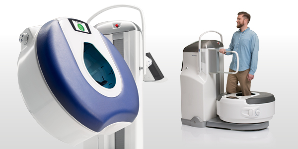
Planmed Verity CBCT unit was developed due to the above-mentioned limitations of MSCT, MRI etc., and based on the requirements of orthopedics and trauma surgery. The CBCT unit provides orthopedists and trauma surgeons with multiplanar cross-sectional information on the extremities and the cervical vertebrae. Users have the option to capture non-weight bearing or natural anatomical weight-bearing images with a high resolution of 0.2 mm in all planes within around 20 seconds. Due to the wide range of applications, the medical CBCT unit has been examined in numerous scientific papers since its first implementation. Its value for orthopedics and trauma surgery in comparison with the commonly available diagnostic methods has been confirmed many times [3 – 6]. Following the requirements of orthopedics and trauma surgery, the radiation hygiene of Planmed Verity is one of the aspects that has been subject to constant development in recent years. As a result, users can now be provided with an Ultra Low Dose (ULD) imaging preset. The resulting radiation dose is lower than that of a digital conventional X-ray in 2 planes typically used in orthopedics and trauma surgery. To develop this ULD preset, the resulting effective dose was determined and compared with the conventional 2D X-ray as well as with MSCT systems typically used in orthopedics and trauma surgery (measurements taken under the same scientific conditions). The results are shown in Figures 1 to 4 for the elbow, wrist, knee joint and ankle joint regions. They confirm that ULD presets enable very high radiation hygiene. The results were able to prove that the resulting effective dose of Planmed Verity is 92.4% lower than that of a MSCT in the wrist region, 91.6% lower in the knee region, 93.5% lower in the ankle region and 97.3% lower in the elbow region. The resulting effective dose is also 31.6% lower compared to a conventional 2D X-ray in the wrist region, 23.3% lower in the knee region, 6.7% lower in the ankle region and 33.3% lower in the elbow region. Planmed Verity was also compared with a conventional 2D X-ray in an isolated study of the wrist region, which took the absorbed dose into account. Here, the absorbed dose of Planmed Verity was 13.2 % lower, while the probability of finding the fracture was 28 % higher on average than with a conventional 2D X-ray. The study thus verified the fundamentally lower dose and the higher value of CBCT imaging for the assessment of osseous structures [7].
Conclusion:
With an option for non-weight bearing or weight-bearing images, a resolution of 0.2 mm and very low exposure times of about 20 seconds, Planmed Verity is optimized for the use in orthopedics and trauma surgery. The higher radiation hygiene of the ULD preset in comparison with the conventional 2D X-ray and with the multi slice CT has been proven in specific measurements which were carried out under scientific standards. Here, the ULD preset with its resulting effective dose was on average 23.73 % lower than with the conventional 2D X-ray, and 93.7 % lower on average than the multi slice CT.
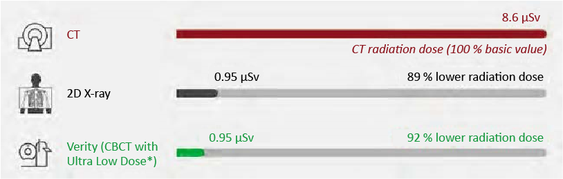
Fig. 1: Representation of the resulting effective radiation dose of a MSCT, a conventional 2D X-ray and CBCT unit using the ULD preset in the wrist region

Fig. 2: Representation of the resulting effective radiation dose of a MSCT, a conventional 2D X-ray and CBCT unit using the ULD preset in the knee joint region

Fig. 3: Representation of the resulting effective radiation dose of a MSCT, a conventional 2D X-ray and CBCT unit using the ULD preset in the ankle region

Fig. 4: Representation of the resulting effective radiation dose of a MSCT, a conventional 2D X-ray and CBCT unit using the ULD preset in the elbow region
Literature
1. Breederveld, R.S., & Tuinebreijer, W. E. (2004). Investigation of computed tomographic scan concurrent criterion validity in doubtful scaphoid fracture of the wrist. Journal of Trauma and Acute Care Surgery, 57 (4), 851 – 854. doi: 10.1097/01.TA.0000124278.29127.42
2. Cruickshank, J., Meakin, A., Breadmore, R., Mitchell, D., Pincus, S., Hughes, T., Bently, B., Harris, M. and Vo, A. (2007), Early computerized tomography accurately determines the presence or absence of scaphoid and other fractures. Emergency Medicine Australasia, 19: 223 – 228. https://doi.org/10.1111/j.1742-6723.2007.00959.x
3. Collan, L, Kankare, JA, Mattila, K. (2013), The biomechanics of the first metatarsal bone in hallux valgus: A preliminary study utilizing a weight bearing extremity CT. Foot Ankle Surg. 19 (3), 155 – 161 http://dx.doi.org/10.1016/j.fas.2013.01.003
4. Burnett, A. L., Chang, A.G., Crone, J.K., Huang, P. L., & Sezen, S. F. (2002). Noncholinergic penile erection in mice lacking the gene for endothelial nitric oxide synthase. Journal of andrology, 23 (1), 92 – 97. https://doi.org/10.1002/j.1939-4640.2002.tb02601.x
5. Colin, F., Horn Lang, T., Zwicky, L., Hintermann, B., & Knupp, M. (2014). Subtalar joint configuration on weightbearing CT scan. Foot & ankle international, 35 (10), 1057 – 1062 doi: 10.1177/1071100714540890.
6. Huang, A. J., Chang, C.Y., Thomas, B. J., MacMahon, P.J., & Palmer, W. E. (2015). Using cone-beam CT as a low-dose 3D imaging technique for the extremities: initial experience in 50 subjects. Skeletal radiology, 44 (6), 797 – 809. https://doi.org/10.1007/s00256-015-2105-9
7. Neubauer, J., Benndorf, M., Ehritt-Braun, C., Reising, K., Yilmaz, T., Klein, C., ... & Goerke, S. M. (2018). Comparison of the diagnostic accuracy of cone beam computed tomography and radiography for scaphoid fractures. Scientific reports, 8 (1), 1-6 https://doi.org/10.1038/s41598-018-22331-8
The effective doses of this study were calculated based on earlier studies for evaluating the doses:
Koivisto, J., Kiljunen, T., Wolff, J., & Kortesniemi, M. (2013). Assessment of effective radiation dose of an extremity CBCT, MSCT and conventional X ray for knee area using MOSFET dosemeters. Radiation protection dosimetry, 157 (4), 515 – 524.
Koivisto, J., Kiljunen, T., Kadesjö, N., Shi, X. Q., & Wolff, J. (2015). Effective radiation dose of a MSCT, two CBCT and one conventional radiography device in the ankle region. Journal of foot and ankle research, 8 (1), 1 – 11.
Koivisto, J., Van Eijnatten, M., Kiljunen, T., Shi, X. Q., & Wolff, J. (2018). Effective radiation dose in the wrist resulting from a radiographic device, two CBCT devices and one MSCT device: a comparative study. Radiation protection dosimetry, 179 (1), 58 – 68.
Koivisto, J., van Eijnatten, M., Ludlow, J., Kiljunen, T., Shi, X. Q., & Wolff, J. (2021). Comparative dosimetry of radiography device, MSCT device and two CBCT devices in the elbow region. Journal of applied clinical medical physics, 22 (5), 128 – 138.